Once
we know what a cataract is, let’s have a look at the Management Of Cataract
First
and foremeost, there are no medicines
which help resolve a cataract. Our prime responsibility is to spread
awareness of this basic fact and protect gullible patients from Quackery and
downright dangerous methods claimed to ‘cure’ the cataract.
Surgery
performed by a trained Ophthalmologist in a sterile Operation Theatre is the Only
safe, approved way to resolve a Cataract
No matter what you do, no matter how hard you try, a scratched windshield needs to be replaced. This is exactly what we do in a Cataract Surgery. We REMOVE the aged/damaged lens; and REPLACE it with an artificial one.
The major strides in the
evolution of Cataract Surgery have been first in the Method of Removal, and subsequently in the Implanted Artificial Lens.
Coaching technique described
in Sushrutha Samhita.
HISTORY
The first interventions for lens of the eye date back
to 2000 B.C. in Egyptian empire and Indian Sciences. Back then, the procedure
was limited to merely displacing the lens from its position within the eye
itself – without any surgery – COUCHING.
This would bring about a mild improvement in vision. The techniques used were
understandably barbaric and rudimentary.
Then came a back and forth change as to whether to
remove the lens with (intracapsular) or
without (extracapsular) the entire Capsule.
The French ophthalmologist Jacques Daviel (1696–1762) was the first modern European physician
to successfully extract cataracts from the eye. He performed the first EXTRACAPSULAR CATARACT EXTRACTION on
April 8, 1747.
Albrech von Graefe (1828-1870), a
tremendously important Ophthalmologist, died at the early age of 42. He gave a
dissertation on 'modified linear extraction' among other significant papers at
the 3rd International Congress of Ophthalmology, Paris. Ophthalmology developed
tremendously through the application of the ophthalmoscope by von Graefe.
At the turn of the Century, a medical student told Sir Nicholas Ridley, that it was a pity
that the cloudy lens extracted could not be replaced with a clear one. At that
time, Sir Nicholas was a surgeon at Moorfield Eye Hospital and St Thomas'
Hospital in London, specialising in Ophthalmology. Dissatisfied with the poor
acuity and loss of binocular single vision following cataract extraction and
the poor outcome, with the contact lenses then available, he had early in his
career envisaged using an artificial lenticulus. He went on to pioneer ARTIFICIAL INTRAOCULAR LENS TRANSPLANTSURGERY for cataract patients.
In 1967, American Charles D. Kelman (1930-2004), an ophthalmologist
pioneer in cataract surgery, introduced PHACOEMULSIFICATION
after being inspired by his dentist's ultrasonic probe. This technique use
ultrasonic waves to emulsifythe lens in order to remove the cataracts without a
large incision. This new method of surgery decreased the need for an extended
hospital stay and made the surgery less painful. The principle behind this has
now been taken to cutting edge technology.
With the development in IOLs, the method of access to the catracted
lens, removing it and Implanting the IOL developed further.
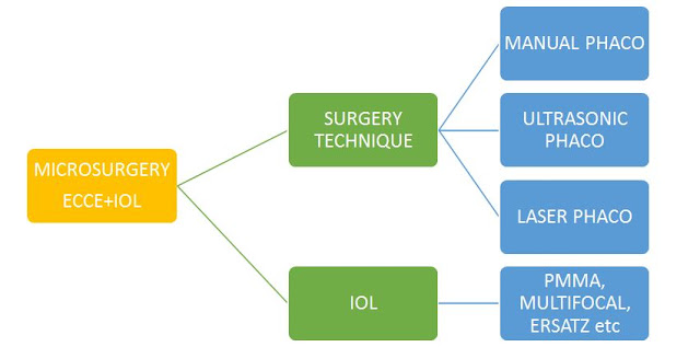
MICROSURGERY MADE COUCHING AND ICCE OBSOLETE
Most
cataract extractions are now ExtraCapsular i.e. Surgeons leave the Posterior
Capsule behind as a base for the artificial Intra Ocular Lens.
MICROSURGERY involves Extracpasular
Lens Extraction and IOL Implantation.
Thanks to recent advances, the Size of Incision required is down from
6-10 mm to 2mm i.e. From an
incision that went halfway round the eye, to barely the tip of a pen.
PHACOEMULSIFICATION
Microscopic surgery and Flodable lenses enabled the
entire procedure to be accomplished through these tiny ports.
The IOL – to be discussed
in subsequent blog post.
Majority
of the cataract surgeries under VisionFoundation of India are performed via Phacoemulsification.
The
Lens implanted is the Indian foldable
lens.
So now that you know about
Phaco, you can reassure prospective patient that :
- · It is Bladeless – a very tiny incision is all that’s needed
- · There are No Stitches – since the incision is small, it self-heals
- · Post op care is simple and in a vast majority, uncomplicated
- · A clear lens is implanted to restore optimum vision
So,
what can you do now?
1.
Spread the Word!
By going through this blog, you’ve
helped already! Next time you come across someone with possible problems with
vision, do not hesitate to reassure them and refer them to us at - VISION
FOUNDATION OF INDIA , 5, Babulnath Road, Mumbai 400 007, India. Tel:
+91-22-23627777 / +91-22-23629999
2.
Donate!
Phaco Cataract Surgeries under Vision Foundation of India
are presently performed at just Rs.
1500 per eye!
3.
Join!
Join the movement as a
Member/Volunteer/Brigadier. Whatever your skillset, VFI needs you. Find out how
you can help. Drop us an email at eyecare@visionfoundationofindia.in
In Ophthalmology, Ignorance is not Bliss – it’s
Blindness.
Next Blog
Article : Preparing for Catarct Surgery
A VFI Youth
Wing Initiative.


No comments:
Post a Comment